In a normal early pregnancy the diameter of the yolk sac should usually be. In patients with accurate dates cardiac motion is seen typically after 5 57 weeks while a yolk sac is first seen at 5 17ths to 5 57ths weeks.

U ltrasound pictures illustrating the measurement of GSD in embryos with CRL of 2 mm left and 25 mm right.
Yolk sac size chart. Yolk sac Imaged 5 - 55 w Imaged when MSD 5-6 mm Imaged 3-5 d prior to embryo Diameter peaks at 6 mm at 10 w then decreases Usually not visible after first trimester Number of yolk sacs usually equals number of amnions. Yolk Sac Size Chart In Cm. Posted on August 1 2019 by Eva.
First trimester of pregnancy gestational sac diameter and yolk hatchability pregnancy from 4 10 weeks imprinted genes. Changes In Gestational And Yolk Sac Diameters All Patients Scientific Diagram. In a normal early pregnancy the diameter of the yolk sac should usually be.
A yolk sac 6 mm is suspicious for a failed pregnancy but not diagnostic. The yolk sac is measured from inner rim to inner rim diameter. Gestational Sac And Yolk Size Chart - Best Picture Of Chart AnyimageOrg.
First trimester of pregnancy defining safe criteria to diagnose gestational sac yolk abnormal pregnancy from 4 10 weeks defining safe criteria to diagnose Normal 1st Trimester Ultrasound How ToEstimating Fetal Gestational Age Radiology KeyNormal 1st Trimester Ultrasound How. Yolk sac YS chart. Corpus luteum CL chart during pregnancy.
Fetal heart rate FHR chart. Crown-rump length CRL chart. Ovulation monitoring during menstrual cycle.
Beta hCG results database. WHO Child Growth Standards. The inner diameter of yolk sac was measured by Transvaginal sonography and its correlation with pregnancy outcome was studied.
The mean yolk sac diameter was noted as 3718 mm. The diameter of the smallest yolk sac was 125 mm and that of the largest was 896 mm. Yolk sac size was normal in 62 8857 cases it was.
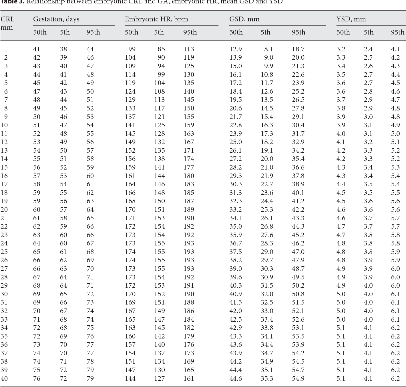
Sion of the yolk sac middle 14 mm. U ltrasound pictures illustrating the measurement of GSD in embryos with CRL of 2 mm left and 25 mm right. The callipers are placed at the inner edges of the trophoblast.
Color version available online Color version available online. When the MSD measures 8 mm a yolk sac should be visible however lack of a yolk sac is not an indication of pregnancy failure. When the MSD measures 25 mm a fetal pole should be visible.
When the MSD measures 20 mm a yolk sac should be visible. Intrauterine gestational sac identification via ultrasonography has 976 specificity for the diagnosis of IUP while the yolk sac visualization has 100 specificity. A normal-appearing gestational sac is smooth round or oval and is in the central portion of the uterus other locations of the gestational sac should raise concerns for ectopic.
The mean Gestational sac diameter. Weeks GS diameter cm 50 10. The gestational sac diameter was 209mm right where it should be.
Ave 174mm range 117mm 5-243mm 95 And the yolk sac diameter was 25mm far far far below what it should be. Ave 36mm range 27mm 5 to 45mm 95. Ovarian Stromal Tumors The size of the yolk sac remains mostly constant until around an embryonic length of 40 centimeters 16 inches whereupon it begins to shrink mostly or completely disappearing by an embryonic length of 50 centimeters 20 inches Eggs should hatch around 20 hours and will survive on the yolk sac for 2 - 3 days at which time they need to be fed microscopic algae.
Feeding the larvae is. An experienced sonographer can detect a yolk sac with transvaginal ultrasound when the gestational sac has reached a mean diameter of 8 mm to 10 mm. The presence of a yolk sac confirms the diagnosis of an intrauterine pregnancy and excludes ectopic pregnancy except in rare cases of simultaneous intrauterine and extrauterine gestations.
Very Large Yolk Sac and Bicornuate Uterus in a Live Birth A case report in which a yolk sac was measured at 81mm but resulted in a live birth. This report states that the quality of the yolk sac might be more important than its size. The yolk sac is the earliest fetal structure that forms in the gestational sac within the uterus during pregnancy.
Having a yolk sac that is too large or too small has been associated with pregnancy loss. However abnormal sac size occurs in approximately 17 of pregnancies. In many cases women go on to have normal pregnancies.
Yolk sac is 48mm - made the sonographer measure it 3 times. Heart rate is 176 and the baby measures 201mm. I have no idea why the yolk sac measured so big last time - the dr said the measurements are tiny and its easy to make a mistake.
All they could see at that point was the gestational sac and yolk sac which were apparently measuring too large measuring at 6 weeks 4 days. The technician told me to prepare myself for a miscarriage or DC. I then had a follow up ultrasound a week later.
There was a fetus and heartbeat measuring at 6 weeks 4 days. The yolk sac is a membranous sac attached to the embryo. It can be seen on ultrasound between the embryo and the gestational sac.
The yolk sac functions as a means for the nourishment of the embryo before the circulatory system and the placenta develop. Measurements of the yolk sacs size and shape are important when assessing the. The fetal crown-rump length CRL is defined as the longest length of the fetus excluding the limbs and yolk sac.
It is the measurement between the top of the head to the area above where the legs begin. The fetal crown-rump length is taken via ultrasound usually up to the 14th week of the pregnancy. This chart shows approximate crown-rump.
Yolk Sac Size Ultrasound I took a little video of a quick ultrasound I had last Wednesday and I go again this Wednesday for my dating ultrasound. She had clicked on what I believe was the yolk sac and in the left corner of the video I can see. Diameter looked to be 409 cm.
This woman didnt do any kind of definitive ultrasound she just kind of looked around quickly confirming pregnancy. Possible Pregnancy Loss. Small gestational sac along with some other early ultrasound findings such as enlarged yolk sac or small gestational sac in relation to the size of embryo measured by crown-rump length may not be enough to definitively diagnose a miscarriage or other pregnancy loss such as a blighted ovum.
By d 36 the embryo is near the top of the vesicle and the yolk sac is all but gone. By d 40 the embryo is back in the middle of the vesicle suspended by the umbilicus. Vesicles that are smaller than normal size for their age are associated with increased rates of embryonic loss.
In patients with accurate dates cardiac motion is seen typically after 5 57 weeks while a yolk sac is first seen at 5 17ths to 5 57ths weeks. When an embryonic heartbeat is first seen the MSD is 20 mm and at the time of embryo movements the MSD reaches 30mm. The yolk sac has an echogenic periphery with a sonolucent center.