04
Apr
IARC 150 Cours Albert Thomas 69372 Lyon CEDEX. This article discusses the common benign cysts found in the vagina and vulva.
Nabothian cyst pathology outlines. A nabothian cyst is a lump filled with mucus on the surface of the cervix or cervical canal. The cervix is located at the lower end of the womb uterus at the top of the vagina. It is about 1 inch 25 centimeters long.
The cervix is lined with glands that normally secrete mucus. Patient age ranged from 20 to 75 years with a mean of 35 years and a peak incidence between 31-40 years 13 cases 325. The majority of patients were asymptomatic 31 cases 775.
The cyst type which was more frequently associated with symptoms was Bartholins duct cyst. Nabothian Cyst of Uterine Cervix is a benign mucus-filled cyst that is present on the cervical wall. It is common tumor found mostly in middle-aged and older women who have had multiple pregnancies.
There are no clearly established risk factors for Nabothian Cyst of Uterine Cervix. The tumor appears benign and can be misdiagnosed as multiple nabothian cysts. 2 This rare variant of adenocarcinoma of the cervix accounts for about 13 of all cervical adenocarcinomas.
2 This case was reported because of the rare incidence of cervical adenoma malignum and the difficulty in making a histopathologic diagnosis because of its. Jobs Fellowships Conferences Cases CME. Uterus Vulva vagina female urethra.
Nabothian Cyst of Uterine Cervix is a benign mucus-filled cyst that is present on the cervical wall. It is common tumor found mostly in middle-aged and older women who have had multiple pregnancies. There are no clearly established risk factors for Nabothian Cyst of Uterine Cervix.
Located at a much higher position within the uterine cervix. Adenoma malignum of the cervix extending to upper vagina. Canal of Nuck hydrocele.
Mucus cysts of vagina. Derived from Mullerian duct. Epithelial inclusion cyst 5.
Atrophic menopausal smear atrophic pattern Smear showing an atrophic pattern taken in a menopausal woman naturally or after treatment. The cytopathologic features of the atrophic cervico-vaginal smear are. A variable cellularity with miniature polygonal to round squamous cells with orangeophilic cytoplasm and dot like pyknotic nuclei red atrophy or pseudoparakeratosis.
Nabothian cysts are tiny cysts that form on garter surface of your cervix. Vaginal pimples can sometimes be a symptom of an underlying condition. These can be caused by injury during childbirth fluid buildup in your glands or benign noncancerous tumors within the vagina.
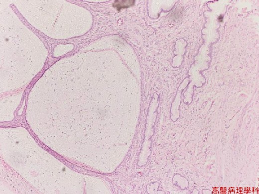
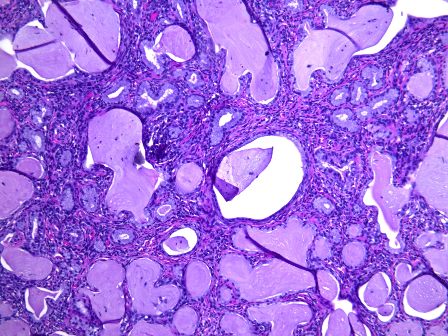
Nabothian cysts also called mucinous retention cysts or epithelial cysts are a common and benign gynecological condition located at the uterine cervix. They are usually asymptomatic of no clinical significance and require no treatment. However if symptomatic or they become complicated further evaluation and therapy are needed.
Pathology Outlines - Nabothian cyst Colloid cysts of the third ventricle are benign epithelial lined cysts with characteristic imaging featuresAlthough usually asymptomatic they can rarely present with acute and profound hydrocephalus. It is a specific type of giant cell in which several epithelioid meaning the cells are big and pink like the cells of the skin macrophages fuse together the nuclei forming a cute horse-shoe shape around the periphery of the cell The topic Cervical Mucinous Retention Cyst you are seeking is a synonym or alternative name or is closely related to the medical condition Nabothian Cyst of Uterine Cervix. Nabothian cysts Squamous metaplasia grows over endocervical glands Mucin gets trapped in glands leading to cystic dilation Nabothian cyst Extremely common of no clinical significance.
Can be very large. Red appearing epithellium is columnarrPink appearing epithelium is squamousrBorder region is transformation zone. Must be distinguished from lymphoma a very rare primitive tumour of uterine cervix.
In cervical smears follicular cervicitis is detected by areas with lymphoid cels of different sizes and in rare zones elements suggesting centro-follicular structures. Histopathology atlas Cytopathology atlas. IARC 150 Cours Albert Thomas 69372 Lyon CEDEX.
This article discusses the common benign cysts found in the vagina and vulva. While not commonly encountered in routine surgical pathology practice these cysts are a relatively frequent occurrence. The cysts are often asymptomatic and do not come to clinical attention.
Cysts of the vagina and vulva can be classified as embryologic or non-embryologic in origin with müllerian and. HIPERPLASIA ENDOMETRIAL ATIPICA PDF. January 23 2021 by admin.
Endometrial hyperplasia EH is a condition in which the innermost lining of the uterus or endometrium undergoes thickening usually as a result of exposure to. Prior to the World Health Organization classified endometrial hyperplasia as simple versus complex and nonatypical. Clear Blue Price Cysts Ovary Pathology Outlines Cppc2013.
Cervical dysplasia Cervical incompetence Cervical polyp Cervicitis Female infertility Cervical stenosis Nabothian cyst Increased concentrations of CEA and CA19-9 in the cyst fluid are described in cystic tumours but not in simple liver cysts. However comparing the levels of CA19. Nabothian cysts are usually considered benign findings and can occur in about 12 of the population Fogel 1982.
Nabothian cyst near external os of cervix arrow. Fogel 1982 Here is an pelvic ultrasound video we made showing you the sagittallongitudinal view. Vulvar squamous hyperplasia Pathology outlines.
Squamous cell hyperplasia previously known as hyperplastic dystrophy or leukoplakia is an excessive growth of normal or abnormal skin in the vulvar region. The condition is thought to be due to chronic irritation. Symptoms of Squamous Cell Hyperplasia Vulvar Pathology - Diagnosis Amie Kawasaki MD Assistant Fellowship Program Director.
Previous post
Nadi astrology jayanagar bangaloreNext post
Naaf ka utarna symptoms